
Fig. 1
- ID
- ZDB-FIG-120720-55
- Publication
- Okuda et al., 2012 - lyve1 expression reveals novel lymphatic vessels and new mechanisms for lymphatic vessel development in zebrafish
- Other Figures
- All Figure Page
- Back to All Figure Page
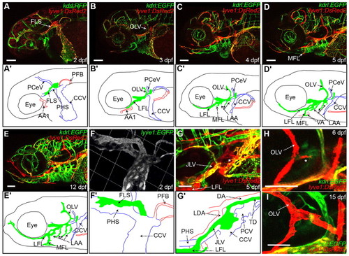
The facial lymphatics originate from the common cardinal vein to form a complex network of vessels. (A-E′) Lateral images of head vessels in the lyve1:EGFP;kdrl:RFP and lyve1:DsRed2;kdrl:EGFP transgenics at 2-12 dpf (A-E), with schematic diagrams of arteries (red), veins (blue) and lymphatics (green) at each stage (A′-E′). (F,F′) 3D reconstruction of the FLS in the lyve1:EGFP transgenic at 2 dpf (F), with schematic diagram (F′′). (G,G′) Dorsolateral image of the JLV and its connection to the TD and the CCV (indicated by a white asterisk) in the lyve1:EGFP;kdrl:RFP transgenic at 5 dpf (G), with schematic diagram (G2). (H,I) Images showing the OLV branching at 6 dpf (H) (indicated by a white asterisk) and connecting to the remnant lyve1-expressing structure attached to the CCV at 15 dpf (12/16) (I) in the lyve1:DsRed2;kdrl:EGFP transgenic. A-E are montage images. A used three z series stacks and B-E used two z series stacks. AA1, mandibular arch; CCV, common cardinal vein; DA, dorsal aorta; FLS, facial lymphatic sprout; JLV, jugular lymphatic vessel; LDA, lateral dorsal aorta; LFL, lateral facial lymphatic; LAA, lymphatic branchial arches; MFL, medial facial lymphatic; OLV, otolithic lymphatic vessel; PCeV, posterior cerebral vein; PCV, posterior cardinal vein; PFB, pectoral fin bud; PHS, primary head sinus; VA, ventral aorta. Scale bars: 100 μm in A-G′; 50 μm in H,I. |