
Fig. S3
- ID
- ZDB-FIG-120720-51
- Publication
- Okuda et al., 2012 - lyve1 expression reveals novel lymphatic vessels and new mechanisms for lymphatic vessel development in zebrafish
- Other Figures
- All Figure Page
- Back to All Figure Page
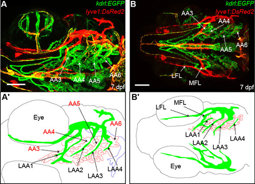
The lymphatic branchial arches develop along the arterial branchial arches. (A,A′) Ventrolateral images of vessels of the pharyngeal area in the lyve1:DsRed2;kdrl:EGFP transgenic at 7 dpf (A), with schematic diagram of arteries (red), veins (blue) and lymphatics (green) (A′). (B,B′) Ventral image of head vessels in the lyve1:DsRed2;kdrl:EGFP transgenic at 7 dpf (B), with schematic diagram (B′). A and B are montage images of two z series stacks. AA3, first branchial arch; AA4, second branchial arch; AA5, third branchial arch; AA6, fourth branchial arch; LAA1, first lymphatic branchial arch; LAA2, second lymphatic branchial arch; LAA3, third lymphatic branchial arch; LAA4, fourth lymphatic branchial arch; LFL, lateral facial lymphatic; MFL, medial facial lymphatic. Scale bars: 100 μm. |