
Fig. S2
- ID
- ZDB-FIG-120720-50
- Publication
- Okuda et al., 2012 - lyve1 expression reveals novel lymphatic vessels and new mechanisms for lymphatic vessel development in zebrafish
- Other Figures
- All Figure Page
- Back to All Figure Page
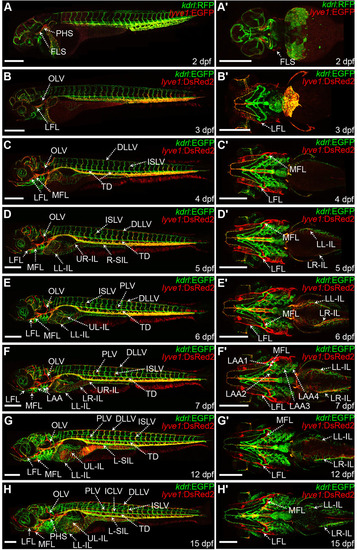
Atlas of lymphatic development in zebrafish from 2 dpf to 15 dpf. (A-H′) Montage of maximum projections of iz series stacks of the entire lyve1:DsRed2;kdrl:EGFP and lyve1:EGFP;kdrl:RFP transgenics (lateral views) at 2-7 dpf (A-F), 12 dpf (G) and 15 dpf (H), and their corresponding ventral images of the head and the anterior intestine (A′-H′). DLLV, dorsal longitudinal lymphatic vessel; ICLV, intercostal lymphatic vessel; ISLV, intersegmental lymphatic vessel; FLS, facial lymphatic sprout; LAA1, first lymphatic branchial arch; LAA2, second lymphatic branchial arch; LAA3, third lymphatic branchial arch; LAA4, fourth lymphatic branchial arch; LFL, lateral facial lymphatic; LL-IL, lower-left intestinal lymphatic; LR-IL, lower-right intestinal lymphatic; L-SIL, left supraintestinal vessel; PHS, primary head sinus; PLV, parachordal lymphatic vessel; MFL, medial facial lymphatic; OLV, otolithic lymphatic vessel; TD, thoracic duct; UL-IL, upper-left intestinal lymphatic; UR-IL, upper-right intestinal lymphatic. Scale bars: 300 μm. |