
Fig. 2
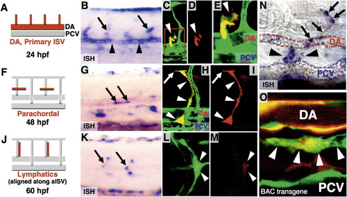
Chemokine Receptor Cxcr4a Expression in the Developing Zebrafish Trunk (A, F, and J) Diagrams indicating Cxcr4a expression domains as red at stages shown below. (B, G, and K) WISH images corresponding to left panels (arrows, Cxcr4a expression domains; arrowheads, Cxcr4a expression in DA at 24 hpf). (C–E) Confocal images of a 35 hpf Tg(fli1a:EGFP)y1 embryo (green) injected with a recombineered BAC clone with TagRFP-T driven by the cxcr4a promoter (red). TagRFP-T is detected in lymphatic progenitors sprouting from the PCV. (C) Merged image. (D) Red channel only. (E) Magnified view in the red box of (C). (H and I) Confocal images of a 45 hpf Tg(fli1a:EGFP)y1 embryo (green) injected with a recombineered BAC clone with TagRFP-T driven by the cxcr4a promoter (red). TagRFP-T is detected in sprouting lymphatic progenitors forming the PC. (H) Merged image. (I) Red channel only. (L and M) Confocal image of a 62 hpf Tg(fli1a:EGFP)y1 embryo (green) injected with a recombineered BAC clone with TagRFP-T driven by the cxcr4a promoter (red). TagRFP-T is detected in LEC migrating dorsally and ventrally from the PC. (L) Merged image. (M) Red channel only. (N) High-magnified WISH image showing a cxcr4a-expressing TD sprout extending along the intersegmental boundary (arrows) and between the DA and PCV (arrowheads) at a 3 dpf animal. (O) Confocal image of ventral trunk vessels in a 5 dpf Tg(fli1a:EGFP)y1 embryo (green) injected with a recombineered BAC clone with TagRFP-T driven by the cxcr4a promoter (red). TagRFP-T expression is present in both TD (arrowheads) and DA, but not in the PCV. See also Figure S2. |
Reprinted from Developmental Cell, 22(4), Cha, Y.R., Fujita, M., Butler, M., Isogai, S., Kochhan, E., Siekmann, A.F., and Weinstein, B.M., Chemokine Signaling Directs Trunk Lymphatic Network Formation along the Preexisting Blood Vasculature, 824-836, Copyright (2012) with permission from Elsevier. Full text @ Dev. Cell