
Fig. 1
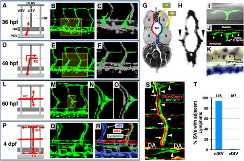
Anatomy and Assembly of the Trunk Lymphatic Network in the Developing Zebrafish (A, D, L, and P) Diagrams illustrating successive steps in trunk lymphatic network assembly at different developmental stages (red, developing lymphatics; red arrows, their growth direction). (B, E, M, and Q) Confocal images of the vasculature in Tg(fli1a:EGFP)y1 animals at each stage (yellow boxes, magnified areas shown to the right). (C, F, N, and O) Magnified views of the boxed portions of the images to the left (green, developing lymphatic vessels, except N). (G) Cross-sectional diagram illustrating lateral growth of lymphatic progenitors (green) toward the superficial HM (red, arterial BVs; blue, venous BVs; No, notochord; NT, neural tube; S, somites). (H) Cross-sectional reconstruction (rotated 90°) of confocal stacks used in (E) (arrowheads, vascular spouts extending laterally along the HM). (I and J) Dorsal view confocal/DIC (I) and confocal only (J) images of a single branching PC sprout (arrowheads) in the superficial HM of a 60 hpf Tg(fli1a:EGFP)y1 larva. (K) Dorsal view image of a branching PC sprout (arrowheads) in the superficial HM of a 56 hpf larva WISH-stained for prox1. (R) Color-coded image of (Q) (green, lymphatic vessels; red, arterial BVs; blue, venous BVs). (S) Confocal image of an aISVs (red/yellow, arrows) with a coaligned ISLV and other developing lymphatics (green, arrowheads) in a 3 dpf Tg(flk:mCherry), Tg(fli1a:EGFP)y1 double transgenic animal. (T) Quantification of aISV and vISV (determined by observing vessels using confocal microscopy at 5 dpf) with a coaligned ISLVs. The total number of aISVs or vISVs counted is shown above each bar. See also Movie S1 and Figure S1. |
Reprinted from Developmental Cell, 22(4), Cha, Y.R., Fujita, M., Butler, M., Isogai, S., Kochhan, E., Siekmann, A.F., and Weinstein, B.M., Chemokine Signaling Directs Trunk Lymphatic Network Formation along the Preexisting Blood Vasculature, 824-836, Copyright (2012) with permission from Elsevier. Full text @ Dev. Cell