
Fig. S7
- ID
- ZDB-FIG-120405-28
- Publication
- Jao et al., 2012 - A zebrafish model of lethal congenital contracture syndrome 1 reveals Gle1 function in spinal neural precursor survival and motor axon arborization
- Other Figures
- All Figure Page
- Back to All Figure Page
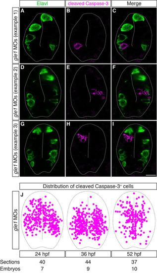
The distribution of cleaved caspase 3+ cells is mutually exclusive with that of differentiated neurons in Gle1-deficient embryos. (A-I) Transverse cryosections through the spinal cords of gle1 morphants were co-stained with anti-Elavl and anti-cleaved caspase 3 antibodies at 24 hpf. The cleaved caspase 3+ cells (B,E,H) are distributed widely along the dorsoventral axis of the spinal cord, often located close to the central canal or immediately adjacent to the basally located Elavl+ neurons (A,D,G). However, cleaved caspase 3+ and Elavl+ signals do not overlap (merged channels, C,F,I). Each image is a single optical section (<1 µm) extracted from the confocal z-stack with dorsal upward. Dashed lines outline spinal cords and central canals. Scale bar: 10 µm. (J) Distribution of the cleaved caspase 3+ cells in the transverse sections of the gle1 morphant spinal cords at 24, 36 and 52 hpf. Single confocal sections were manually superimposed and the positions of the cleaved caspase 3+ cells were marked. The cleaved caspase 3+ cells are distributed widely along both the dorsoventral and apical-basal axes of the spinal cord. |