
Fig. 2
- ID
- ZDB-FIG-120405-16
- Publication
- Jao et al., 2012 - A zebrafish model of lethal congenital contracture syndrome 1 reveals Gle1 function in spinal neural precursor survival and motor axon arborization
- Other Figures
- All Figure Page
- Back to All Figure Page
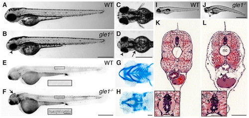
Morphological phenotypes of gle1–/– mutants. (A-D) Lateral (A,B) and dorsal (C,D) views of wild-type (A,C) and gle1–/– (B,D) zebrafish embryos at 2 dpf. Arrowheads (B,D) mark the smaller head and eyes; arrow (D) marks underdeveloped pectoral fins. (E,F) Acridine Orange-stained 2 dpf wild-type (E) and gle1–/– (F) embryos. Cell death is noted in the head (F, arrow) and spinal cord (F, inset). (G,H) Alcian Blue-stained head cartilages of 5-dpf wild-type (G) and gle1–/– (H) larvae. gle1–/– mutant lacks most viscerocranium but has relatively intact neurocranium. (I,J) Lateral views of 5-dpf wild-type (I) and gle1–/– (J) larvae. Asterisk (J) marks pericardial edema. (K,L) Hematoxylin and Eosin (H&E)-stained transverse sections of wild-type (K) and gle1–/– (L) larvae at 5 dpf. gle1–/– mutant has a smaller spinal cord (L, arrow in inset). Asterisk (L) marks subcutaneous edema; arrowhead (L) marks unfolded intestine. nc, notochord; sc, spinal cord. Scale bars: 500 μm in B,D,F,J; 100 μm in H,L and F inset; 50 μm in L inset. |
| Fish: | |
|---|---|
| Observed In: | |
| Stage Range: | Long-pec to Day 5 |