
Fig. 1
- ID
- ZDB-FIG-111115-11
- Publication
- O'Brien et al., 2011 - Wt1a, Foxc1a, and the Notch mediator Rbpj physically interact and regulate the formation of podocytes in zebrafish
- Other Figures
- All Figure Page
- Back to All Figure Page
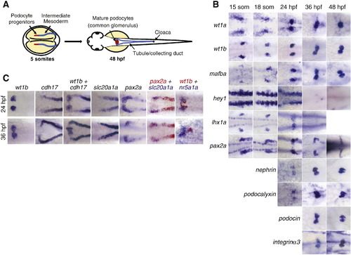
Characterization of podocyte development in zebrafish embryos. A) Schematic of pronephric development in zebrafish. B) Micrographs of in situ hybridizations show the expression patterns of early (wt1a, wt1b, mafba, hey1, lhx1a, pax2a) and mature (nephrin, podocin, podocalyxin, integrinα3) podocyte markers from 15 somites through 48 h of development. C) Analysis of podocyte development in relation to the surrounding tissues. Micrographs of in situ hybridizations compare podocyte (wt1b+, pax2a+ at 24 hpf), neck (pax2a+, wt1b-), first proximal tubule segment (slc20a1a+) and interrenal (nr5a1a+) development at 24 hpf and 36 hpf. Cdh17+ cells represent all non-podocyte renal epithelia which includes both neck and tubule fates. |
| Genes: | |
|---|---|
| Fish: | |
| Anatomical Terms: | |
| Stage Range: | 14-19 somites to Long-pec |
Reprinted from Developmental Biology, 358(2), O'Brien, L.L., Grimaldi, M., Kostun, Z., Wingert, R.A., Selleck, R., and Davidson, A.J., Wt1a, Foxc1a, and the Notch mediator Rbpj physically interact and regulate the formation of podocytes in zebrafish, 318-30, Copyright (2011) with permission from Elsevier. Full text @ Dev. Biol.