
Fig. 7
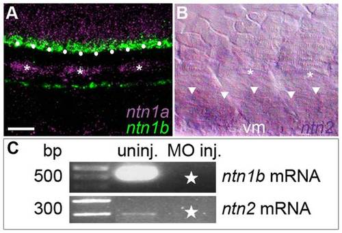
Netrin mRNAs are expressed in intermediate targets of the CaP axon and wildtype transcripts were knocked down with SBMOs. A. Projected confocal stack of ntn1a and ntn1b mRNA expression in zebrafish trunk at 24 hpf; ntn1a is expressed in spinal cord (purple) and muscle pioneers (purple and *). ntn1a staining was false colored purple. ntn1b mRNA (green) is expressed in floor plate (white dots) and hypochord, located ventrally to the notochord. B. Brightfield image of lateral surface of zebrafish trunk at 24 hpf reveals ntn2 is expressed in ventral somite (white arrowheads). Asterisks denote muscle pioneers, cells that do not express ntn2. C. RT-PCR results confirming uninjected embryos have wildtype ntn1b (500 bp band) and ntn2 (300 bp band) transcript. Starred lanes show netrin transcripts are reduced or absent after injecting 10.0 ng of ntn1b SBMO or 5.0 ng of ntn2 SBMO. Multiple samples were run in the gels for ntn1b and ntn2. Lanes containing samples unrelated to the netrins were cropped from the figure. Scale bar = 20 μm. |
| Genes: | |
|---|---|
| Fish: | |
| Anatomical Terms: | |
| Stage: | Prim-5 |