
Fig. 2
- ID
- ZDB-FIG-110922-5
- Publication
- Pyati et al., 2011 - p63 Mediates an Apoptotic Response to Pharmacological and Disease-Related ER Stress in the Developing Epidermis
- Other Figures
- All Figure Page
- Back to All Figure Page
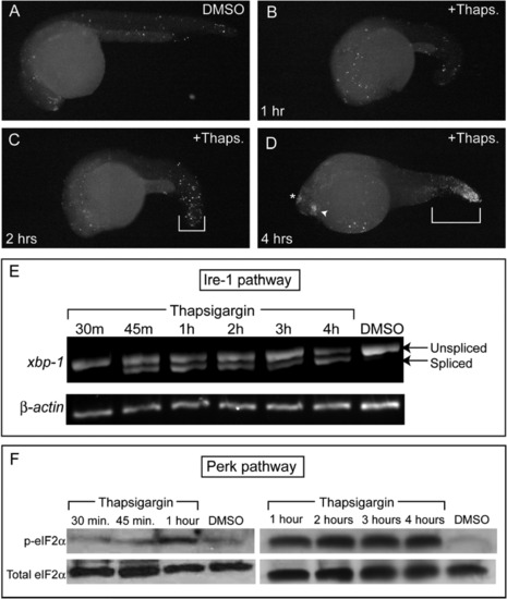
Thapsigargin Treatment Rapidly Activates Conserved Vertebrate ER Stress Pathways to Induce Apoptosis (A–D) TUNEL staining was performed in fixed embryos after (A) 4 hr of DMSO treatment or after (B) 1 hr, (C) 2 hr, or (D) 4 hr of thapsigargin treatment. (C) Increased apoptosis was first apparent in the tail by 2 hr of thapsigargin treatment, but robust apoptosis in the lens, epiphysis, and tail was evident only after (D) 4 hr of thapsigargin treatment compared to DMSO controls at the same stage. All images are representative of embryos examined at each time point (n = 10 in all treatments except D, where n = 9). (E) By RT-PCR, xbp-1 splicing in the Ire-1 pathway was first apparent after 45 min of thapsigargin treatment, and was maintained over 4 hr. Note the lower band present from 45 min on, which corresponds to the nonconventional splice-form downstream of Ire-1 activation. (F) Antiphospho eIF2α immunoblots showed that eIF2α was robustly phosphorylated in the Perk pathway from 1 hr through the end of the thapsigargin time-course. Total eIF2α was immunoblotted as a loading control after stripping of the original blot. |
Reprinted from Developmental Cell, 21(3), Pyati, U.J., Gjini, E., Carbonneau, S., Lee, J.S., Guo, F., Jette, C.A., Kelsell, D.P., and Look, A.T., p63 Mediates an Apoptotic Response to Pharmacological and Disease-Related ER Stress in the Developing Epidermis, 492-505, Copyright (2011) with permission from Elsevier. Full text @ Dev. Cell