
Fig. S2
- ID
- ZDB-FIG-110922-11
- Publication
- Pyati et al., 2011 - p63 Mediates an Apoptotic Response to Pharmacological and Disease-Related ER Stress in the Developing Epidermis
- Other Figures
- All Figure Page
- Back to All Figure Page
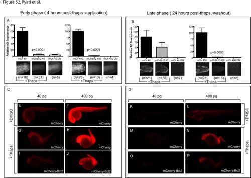
related to Figure 4: Bcl-2 overexpression in zebrafish embryos blocks the early and late apoptotic response after thapsigargin treatment. Embryos injected with mRNAs encoding mCherry or mCherry-Bcl-2 were treated with thapsigargin to induce an early (A, C) or late (B, D) apoptotic response which was analyzed by AO staining. (A) Cropped photos were quantitated for total fluorescence intensity with Volocity software, and values were normalized to controls. Thapsigargin treatment induces early phase apoptosis (A) and late phase apoptosis (B) in embryos injected with 40 or 400 pg mCherry mRNA (mCh 40 or mCh 400 respectively) but not in embryos injected with 40 or 400 pg mCherry-Bcl-2 mRNA (mChBcl2 40 or mChBcl2 400 respectively). No early-phase apoptosis (A) and late-phase apoptosis (B) was observed in embryos injected with 40 or 400 pg mCherry mRNA and treated with DMSO. (C) After 4 hours post thapsigargin treatment, Cherry fluorescence expression, visualized by microscopy, is similar in embryos treated with DMSO (E, F) or thapsigargin (G, H, I, J) and injected either with mCherry alone (E, F, G, H) or with mCherry-Bcl-2 (I, J), for both 40pg and 400 pg concentrations respectively. (D) After 24 hours post thapsigargin treatment, mCherry fluorescence expression, visualized by microscopy, is very weak in embryos injected with 40pg of either mCherry (K, M) or mCherry-Bcl-2 mRNA (O) for both DMSO (K) and thapsigargin treated (M, O) groups. mCherry fluorescence expression is detectable and similar in embryos injected with 400pg (L, N, P) of either mCherry (L, N) or mCherry-Bcl-2 mRNA (P) for both DMSO (L) and thapsigargin treated groups (N, P). |
Reprinted from Developmental Cell, 21(3), Pyati, U.J., Gjini, E., Carbonneau, S., Lee, J.S., Guo, F., Jette, C.A., Kelsell, D.P., and Look, A.T., p63 Mediates an Apoptotic Response to Pharmacological and Disease-Related ER Stress in the Developing Epidermis, 492-505, Copyright (2011) with permission from Elsevier. Full text @ Dev. Cell