
Fig. 1
- ID
- ZDB-FIG-110922-4
- Publication
- Pyati et al., 2011 - p63 Mediates an Apoptotic Response to Pharmacological and Disease-Related ER Stress in the Developing Epidermis
- Other Figures
- All Figure Page
- Back to All Figure Page
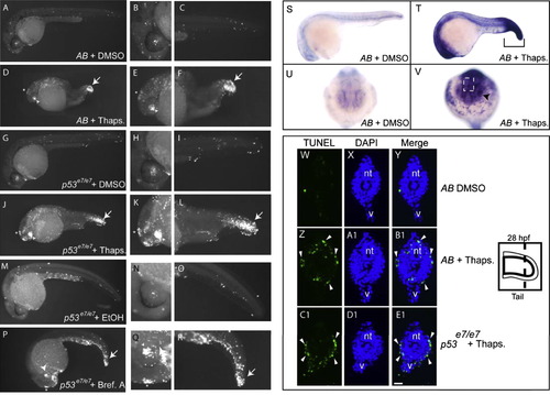
ER Stressors Trigger bip Upregulation and Apoptosis in the Lens, Epiphysis, and Tail Epidermis of 24 Hpf Zebrafish Embryos Embryos (24 hpf) from an AB incross or a stage-matched p53 homozygous mutant incross were treated with a DMSO vehicle or 5 μM thapsigargin for 4 hr, fixed, and assayed for apoptosis by TUNEL labeling. (A–C) Scattered cells in AB DMSO-treated control embryos (n = 29) showed TUNEL positivity. (D–F) Thapsigargin-treated AB embryos had increased TUNEL positivity that was concentrated in the epiphysis, lens, and tail (93% of embryos with increased apoptosis, n = 30). (G–I) p53 homozygous mutant embryos treated with DMSO (n = 31) showed comparable levels of TUNEL positivity to AB DMSO-treated embryos. (J–E1) p53 homozygous mutant embryos treated with thapsigargin showed increased apoptosis in the same regions as in AB embryos (100% of embryos with increased apoptosis, n = 31). Like thapsigargin treatment, Brefeldin A treatment (5 ug/ml) caused increased apoptosis in the lens, epiphysis, and tail (P–R) compared to vehicle-treated controls (M–O) (see also Figure S1). Compared to DMSO-treated controls (S and U), bip expression was upregulated in embryos treated for 3 hr with thapsigargin (T and V). In cryosections, DMSO controls (W–Y) had minimal apoptosis, while thapsigargin treatment of both AB embryos (Z–B1) and p53 mutant embryos (C1–E1) caused extensive enriched apoptosis in the tail epidermis (white arrowheads). In (A)–(R), zoomed images of head and tail are shown at the right of corresponding whole-embryo images. Asterisks indicate epiphysis, arrowheads the lens, and arrows the tail in thapsigargin-treated embryos. |
| Gene: | |
|---|---|
| Fish: | |
| Condition: | |
| Anatomical Terms: | |
| Stage: | Prim-5 |
| Fish: | |
|---|---|
| Conditions: | |
| Observed In: | |
| Stage: | Prim-5 |
Reprinted from Developmental Cell, 21(3), Pyati, U.J., Gjini, E., Carbonneau, S., Lee, J.S., Guo, F., Jette, C.A., Kelsell, D.P., and Look, A.T., p63 Mediates an Apoptotic Response to Pharmacological and Disease-Related ER Stress in the Developing Epidermis, 492-505, Copyright (2011) with permission from Elsevier. Full text @ Dev. Cell