
Fig. S7
- ID
- ZDB-FIG-110922-16
- Publication
- Pyati et al., 2011 - p63 Mediates an Apoptotic Response to Pharmacological and Disease-Related ER Stress in the Developing Epidermis
- Other Figures
- All Figure Page
- Back to All Figure Page
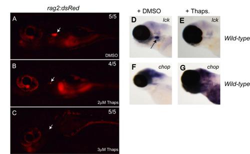
related to Figure 4: Thapsigargin treatment leads to a loss of thymocytes in 5 day old zebrafish embryos. Using the thymocyte specific reporter line, Tg(rag2:dsRed), we observed that thapsigargin treatment leads to a loss of thymocytes in 5 day old embryos as depicted in B (2μM thapsigargin treatment) and C (3μM thapsigargin treatment) as compared with DMSO treated control embryos (A). This phenotype is accompanied by decrease in lck expression in thapsigargin treated embryos (E) compared to DMSO treated control embryos (D) and an increase in chop levels in embryos treated with thapsigargin (G) compared to DMSO treated embryos (F). (white arrows in A,B and C, and black arrow in D show thymus) |
Reprinted from Developmental Cell, 21(3), Pyati, U.J., Gjini, E., Carbonneau, S., Lee, J.S., Guo, F., Jette, C.A., Kelsell, D.P., and Look, A.T., p63 Mediates an Apoptotic Response to Pharmacological and Disease-Related ER Stress in the Developing Epidermis, 492-505, Copyright (2011) with permission from Elsevier. Full text @ Dev. Cell