
Fig. 6
- ID
- ZDB-FIG-110712-17
- Publication
- Chapouton et al., 2011 - Expression of Hairy/enhancer of split genes in neural progenitors and neurogenesis domains of the adult zebrafish brain
- Other Figures
- All Figure Page
- Back to All Figure Page
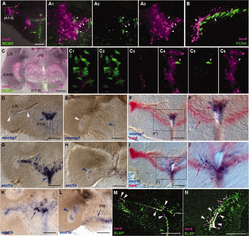
Expression of her genes in the adult hypothalamus characterizes radial glia as opposed to actively dividing neurogenic domains. her Gene expression was detected by using in situ hybridization on 80-μm vibratome cross-sections at levels equivalent to Figure 5B (for A–A3,D,F,G,I,K,M) and 5J (for B–C6,E,H,L,N). A–C6: Comparison of the expression of her4 and her9 (magenta) with the proliferation markers MCM5 or PCNA (green; as indicated) analyzed under confocal microscopy revealed that most domains expressing her genes (magenta arrows) are largely nonproliferating (green arrows). Upon close examination, a few cells coexpressing both marker types can be seen (white arrowheads). A1–3 and C1–6 are high magnifications of the areas boxed in A and C, respectively. B is a high magnification of the LR. D–L: Comparison of the expression of her4 (red) with the proneural genes neurog1, ascl1a, and ascl1b (blue, as indicated) shows apparent coexpression along the medial aspect of the DiV (see F2,I2, purple arrowheads), whereas the patterns diverge more laterally (I2, blue and red arrows). Note the complementary patterns of neurog1 and ascl1 expression dorsal and ventral to the branching point of the LR, respectively. neurog1 Tentatively labels the PTN. Note also the prominent expression of her4 along the LR, contrasting with the weak or absent transcription of proneural genes. Here arrows point to expression along the medial aspect of the LR or the PR, and arrowheads point to the tip of the LR and the DiV. White arrowheads indicate ventricular zones of nonexpression. F2 and I2 are high magnifications of the boxed areas in F and I, respectively. M,N: Costaining for her4 (magenta) and the radial glia protein BLBP (immunocytochemistry, green) showed strong coexpression of the two markers all along the hypothalamic ventricles in confocal analysis (white arrowheads). Some BLBP-positive, her4-negative cells were observed (green arrowheads). LR, lateral recess; PR, posterior recess; PTN, posterior tuberal nucleus. Scale bars = 100 μm. |
| Genes: | |
|---|---|
| Antibody: | |
| Fish: | |
| Anatomical Terms: | |
| Stage: | Adult |