
Fig. 1
- ID
- ZDB-FIG-110712-13
- Publication
- Chapouton et al., 2011 - Expression of Hairy/enhancer of split genes in neural progenitors and neurogenesis domains of the adult zebrafish brain
- Other Figures
- All Figure Page
- Back to All Figure Page
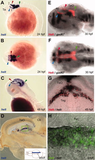
Costaining of embryos and adults for helt mRNA and Helt protein expression demonstrates the specificity of the anti-Helt antibody. A–C: Expression of helt revealed by in situ hybridization on whole-mount embryos (A,C: sagittal views, B: dorsal view; all embryos oriented anterior left, stages indicated). Major expression is detected in the tegmentum. Green arrows point to the posterior expression limit at the midbrain–hindbrain boundary, and red and blue arrows point to anterior expression limits of two tegmental stripes along the borders of pretectum and zona limitans intrathalamica, respectively. This expression is in agreement with the profile described for mouse Helt (Miyoshi et al.,2004; Guimera et al.,2006a; Nakatani et al.,2007). D: Expression of helt revealed by in situ hybridization on a parasagittal section of the adult brain (see cartoon). The midbrain area is shown. E–H: Expression of Helt revealed by the 4A8 antibody (red or green staining) compared with in situ hybridization (black staining) for helt (G,H) or gad67 (E,F) used as landmark. On whole-mount embryos (E,F), note the similar profile of 4A8 with helt mRNA (A,B). G: Cross-section of a 48-hpf embryo at posterior midbrain levels (position indicated by the green arrowhead on C). Note that helt mRNA (black) and immunostainings are found in the same cells. H: Sagittal section of an adult brain (position boxed in D). Again, note the coincidence of helt expression with the 4A8 immunostaining. |
| Genes: | |
|---|---|
| Antibody: | |
| Fish: | |
| Anatomical Terms: | |
| Stage Range: | Prim-5 to Adult |