
Fig. 3
- ID
- ZDB-FIG-110624-8
- Publication
- Ikenaga et al., 2011 - Formation of the spinal network in zebrafish determined by domain-specific pax genes
- Other Figures
- All Figure Page
- Back to All Figure Page
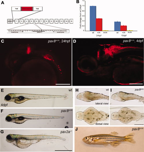
Homozygous embryos are Pax8 hypomorphs. A: The exon composition of zebrafish pax8 indicating the insertion site of Tol2. Primers for the qPCR were designed so that the amplicon spans the junction between exon 5 and exon 6. B: qPCR of the pax8 transcript in wild-type (wt), pax8+/m, and pax8m/m fish at 1 dpf and 10 dpf. Wild type at 1 dpf was used as a reference sample to obtain relative transcript amounts. Error bars represent the SEM of the average from triplicates of a single run (n = 3). C,D: Confocal images of RFP signals in pax8m/m fish at 24 hpf (C) and 4 dpf (D). E–G: The gross morphology of larva at 4 dpf. E is wild type, F is pax8m/m, and G is pax2a–/–. The pax2a–/– larva is deformed and dies at 7–10 dpf. H,I: The brain morphology is normal in the pax8m/m larva (I) compared with the WT larva (H). Top is the lateral view and bottom is the dorsal view. J: Adult pax8m/m fish. Scale bars = 200 μm in C,D; 1 mm in G (applies to E–G; 500 μm in I (applies to H,I). |