
FIGURE
Fig. s1
- ID
- ZDB-FIG-110624-15
- Publication
- Ikenaga et al., 2011 - Formation of the spinal network in zebrafish determined by domain-specific pax genes
- Other Figures
- All Figure Page
- Back to All Figure Page
Fig. s1
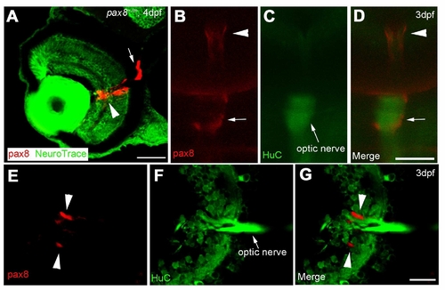
The expression of RFP in glial cells surrounding the optic nerve. A: The RFP signal detected in the eye of a 4 dpf embryo counterstained with NeuroTrace. RFP signal is located around the optic nerve. Scale: 40 μm. B, C, D: In vivo imaging of RFP (B) in a HuC-Cameleon background . Optic nerve emits fluorescence due to the presence of Cameleon (C). D is a merge. Scale: 50 μm. E, F, G: Slice of an eye from HuC-Cameleon fish expressing RFP. RFP (+) cells (E) do not show the expression of Cameleon (F). G is a merge. Scale: 20 7mu;m. Magenta/Green images are provided in Suppl. figure. 7. |
Expression Data
Expression Detail
Antibody Labeling
Phenotype Data
Phenotype Detail
Acknowledgments
This image is the copyrighted work of the attributed author or publisher, and
ZFIN has permission only to display this image to its users.
Additional permissions should be obtained from the applicable author or publisher of the image.
Full text @ J. Comp. Neurol.