
Fig. 2
- ID
- ZDB-FIG-110624-7
- Publication
- Ikenaga et al., 2011 - Formation of the spinal network in zebrafish determined by domain-specific pax genes
- Other Figures
- All Figure Page
- Back to All Figure Page
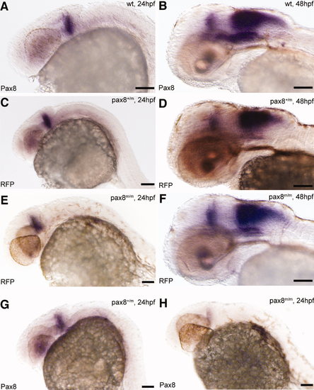
Pax8 expression is identical to RFP expression. Transcripts were detected using in situ hybidization with either pax8 (A,B,G,H) or RFP (C–F) probes in 24-hpf (A,C,E,G,H) or 48-hpf (B,D,F) embryos. Expression pattern, at 24 hpf, of pax8 in wild type (pax8+/+; A) was the same as that of RFP in pax8+/m (C) and pax8m/m (E). Signal was detected in the mhb region of the embryos. RFP showed no signal in wild-type samples (data not shown). At 48 hpf, the staining pattern of pax8 in wild type (B) resembled that of RFP expression in pax8+/m (D) and pax8m/m (F) embryos. Pax8+/m embryos showed positive staining also with the pax8 probe (G). Pax8 transcripts were also detected, at a much lower intensity, in pax8m/m embryos (H). Scale bars = 100 μm. |