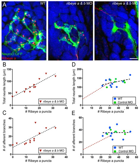
Depletion of Ribeye results in reduced afferent innervation. (A) Representative confocal z-projections showing GFP-labeled innervating afferent neurons (green), Ribeye a immunolabel (red) and DAPI label (blue) in lateral line neuromast 2 from three individual 4 dpf larva. Scale bars: 3 µm. (B,C) Plots of the relationship between either the total length of innervating afferent processes (B) or the number of afferent branch junctions (C) and the number of Ribeye a immunolabeled punctae in ribeye a and ribeye b morphants. Each point represents neuromast 2 in an individual larva. (D,E) Plot of the relationships between the total length of innervating afferent processes (D) or the number of afferent branch junctions (E) and the number of Ribeye a immunolabeled punctae in wild-type and control MO-injected larvae. There was no significant correlation between dendritic length (Pearson′s r=0.4525; P=0.08) or the number of afferent branches (Pearson′s r=0.0129; P=0.97) and the number of Ribeye a punctae. The linear regressions of the plots in either B or C are included for comparison (broken red line).
|
