
Fig. s1
- ID
- ZDB-FIG-110324-35
- Publication
- Sheets et al., 2011 - Ribeye is required for presynaptic CaV1.3a channel localization and afferent innervation of sensory hair cells
- Other Figures
- All Figure Page
- Back to All Figure Page
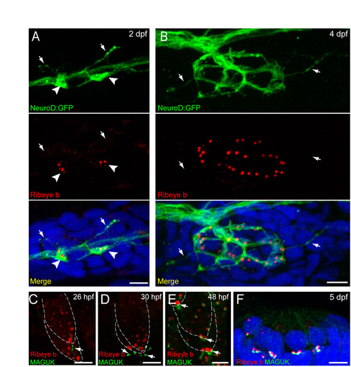
Fig. S1. Afferent innervation and postsynaptic density localization in relation to Ribeye during zebrafish hair-cell development. Scale bars: 3 μm. (A,B) GFP-labeled innervating afferent neurons (green) and Ribeye b immunolabel (red) in a lateral-line neuromast from 2 dpf (A) and 4 dpf (B) larvae. White arrowheads in A indicate dendritic varicosities adjacent to Ribeye-positive aggregates. White arrows in A,B indicate fine dendritic processes that are not adjacent to Ribeye immunolabel. Merged images include DAPI. (C-E) Ribeye b (red) and pan-MAGUK (green) immunolabel in the developing ear at 26 hpf (C), 30 hpf (D) and 48 hpf (E). MAGUK (white arrows) is initially visible at 26 hpf, is beneath but not juxtaposed to Ribeye label at 30 hpf and is adjacent to Ribeye punctae at 48 hpf. (F) Ribeye b (red) and pan-MAGUK (green) labeling in a cross-section of a neuromast at 5 dpf. MAGUK immunolabel is juxtaposed to Ribeye labeled punctae. |
| Genes: | |
|---|---|
| Antibodies: | |
| Fish: | |
| Anatomical Terms: | |
| Stage Range: | Prim-5 to Day 5 |