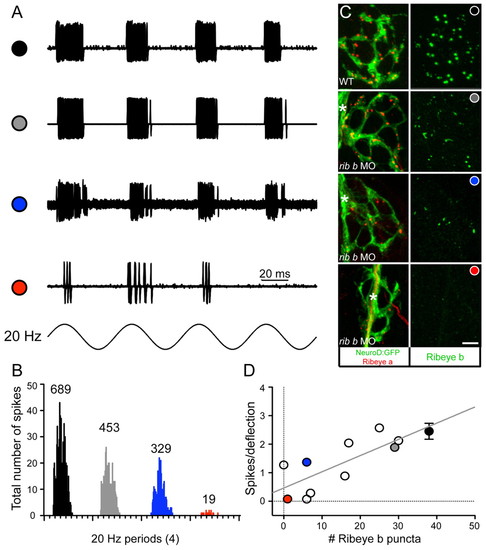
Depletion of Ribeye b results in a reduction in the number of spikes in afferent neurons. (A) Extracellular action currents recorded from a posterior lateral line neuron in response to 20 Hz mechanical stimulation of neuromast hair cells (4 dpf). Shown are overlays of 60 consecutive sweeps from wild-type (black circle) and three ribeye b MO-injected larvae with variable degrees of knockdown (see Results; grey circle, blue circle and red circle). (B) Four frequency histograms (20 Hz) for all spikes recorded in A. Numbers above each histogram indicate the total number of spikes. The overall width and shape of each histogram illustrates that the degree of phase-locking with the stimulus was maintained despite the reduction in spike number. (C) Confocal z-projections of neuromasts from a representative wild-type larva and from the three neuromasts recorded from the ribeye b MO-injected larvae in A. The left panels show Ribeye a staining (red) and the innervating afferent neuron labeled with GFP (green). The right panels show corresponding Ribeye b staining (green). In a larva that was responsive to acoustic stimuli yet displayed a mild balance defect (grey circle), Ribeye b levels were less depleted than the two larvae unresponsive to acoustic stimuli (blue and red circles). Scale bar: 3 μm. Asterisks indicate the tract of the lateral-line nerve. (D) Plot of the relationship between spikes per deflection and the number of Ribeye b punctae. Each circle represents a recorded neuromast from an individual morphant larva, except for the black circle, which represents the average of several wild-type larvae, including that shown in A. The grey, blue and red circles are from the three individual neuromasts in A-C. Error bars indicate s.e.m.
|
