
Fig. 2
- ID
- ZDB-FIG-110218-3
- Publication
- Barrios et al., 2003 - Eph/Ephrin signaling regulates the mesenchymal-to-epithelial transition of the paraxial mesoderm during somite morphogenesis
- Other Figures
- All Figure Page
- Back to All Figure Page
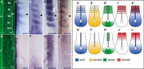
Expression of Eph Family Members in the Paraxial Mesoderm of Wild-Type and fss-/- Embryos Dorsal views of the paraxial mesoderm of 8-somite-stage wild-type and 10-somite-stage fss-/- embryos and schematics with anterior oriented toward the top. The arrowheads indicate the position of the most recently formed intersomitic boundary. (A and B) Living wild-type and fss-/- embryos labeled with Bodipy ceramide. In the wild-type embryo, the positions of the last two somites formed (SII, SI), the forming somite (S0), and the two presumptive somites in the PSM (S-I, S-II) are indicated. The arrowhead points to the intersomitic boundary. (C–J) Expression of (C and D) ephA4, (E and F) ephrin-B2b, (G and H) ephrin-B2a, and (I and J) ephrin-A1 in wild-type (top row) and fss mutant (bottom row) embryos in the region of the paraxial mesoderm shown in (A) and (B). (C′–J′, K, and L) Schematics summarizing expression of Eph family members in the paraxial mesoderm of wild-type and fss-/- embryos. nt, neural tube; n, notochord. The scale bars represent 30 μm. |
| Genes: | |
|---|---|
| Fish: | |
| Anatomical Terms: | |
| Stage: | 10-13 somites |
| Fish: | |
|---|---|
| Observed In: | |
| Stage: | 10-13 somites |