
Fig. 1
- ID
- ZDB-FIG-110218-2
- Publication
- Barrios et al., 2003 - Eph/Ephrin signaling regulates the mesenchymal-to-epithelial transition of the paraxial mesoderm during somite morphogenesis
- Other Figures
- All Figure Page
- Back to All Figure Page
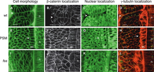
Cells Undergo Mesenchymal-to-Epithelial Transition at Somite Boundaries (A–L) Dorsal views of the left-sided paraxial mesoderm of embryos labeled with Bodipy ceramide (which reveals cell morphology; [A], [E], and [I]) or with Bodipy 505-515 (which reveals nuclear position, [C], [G], and [K]) or immunostained for β-catenin ([B], [F], and [J]) or for γ-tubulin (which labels centrosomes) and stained with phalloidin (which labels actin) ([D], [H], and [L]). Anterior is oriented toward the top. (A–D) Cells at somite boundaries in wild-type embryos. The arrowheads point to the intersomitic boundary. (E–H) Cells in the presomitic mesoderm (PSM) of wild-type embryos. The arrows point to epithelial adaxial cells in which centrosomes are apically localized (H), as also seen in epithelial cells at somite boundaries (D). Centrosomes are randomly positioned in other PSM cells. (I–L) Cells in the somitic mesoderm of fss-/- embryos. n, notochord; ac, adaxial cells. The scale bars represent 10 μm. |