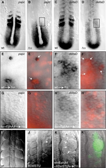
EphA4 Is Unlikely to Induce Boundary Formation by Restoring “Anterior Segmental Identity” to Donor Cells
(A–D) Expression of (A and B) papc and (C and D) deltaD in the paraxial mesoderm of 11-somite-stage (A and C) wild-type and (B and D) fss-/- embryos; anterior is oriented toward the top. Expression in the anterior region of the prospective somites is lost for papc and is reduced for deltaD in fss mutants. The rectangles indicate the areas of the paraxial mesoderm of fss-/- embryos where transplanted cells are shown in (E)–(H).
(E, F, E′, and F′) (E and F) DIC and (E′ and F′) DIC and fluorescent overlays of the paraxial mesoderm of fss-/- embryos in which wildtype cells containing rhodamine dextran (red) were transplanted. The arrows indicate restoration of expression of (E′) papc and high levels of (F′) deltaD expression in transplanted wild-type cells. These data show that the wild-type Fss protein is sufficient to cell-autonomously induce papc and high levels of deltaD expression in wild-type cells within fss-/- hosts. Despite expression of these markers of “anterior segmental identity” in donor cells, ephA4 expression is absent (see Figure 3C in the main text) and boundaries are not induced in the somitic mesoderm (see Figure 3B in the main text).
(G, H,G′, and H′) (Gand H) DIC and (G′ and H′) DIC and fluorescent overlays of the paraxial mesoderm of fss-/- embryos in which EphA4- expressing fss-/- cells containing rhodamine dextran (red) were transplanted. (G′ and H′) Expression of papc or enhanced deltaD expression is not observed in EphA4-expressing fss-/- cells. Therefore, restoration of EphA4 activity in fss-/- cells has no obvious effect on papc or deltaD expression. Despite the absence of these markers of “anterior segmental identity” in donor cells, boundaries are induced in the paraxial mesoderm (see Figure 3D in the main text). These data suggest that EphA4 is acting downstream of the events that specify anterior character in the PSM.
(I and J) Dorsal views of the paraxial mesoderm of a (I) wild-type and a (J) dominant-negative XDeltaSTU-injected embryo. The antimorphic Delta construct is able to disrupt somite boundary formation (asterisk; [S4]). (K and K′) (K) DIC and (K′) DIC and fluorescent overlay of the paraxial mesoderm of an fss-/- embryo where wild-type cells expressing EphA4 and XDeltaSTU (green) have been transplanted. The arrows point to the morphologically distinct boundary at the interface between donor and host cells (seen in nine of ten cases). In fss-/- cells, expression of Notch pathway genes is disrupted [S5] and the XDeltaSTU construct is likely to perturb Notch pathway signaling in the transplanted wild-type cells. In this experiment, Eph/Ephrin signaling still induces boundary formation despite the likely disruption to Notch-Delta signaling (implying that Eph/Ephrin signaling is unlikely to induce boundaries by restoring Notch/Delta signaling interfaces).
From these results, we conclude that the apposition of ephA4-expressing cells and ephrin-expressing cells is sufficient to restore morphologically evident boundaries without requiring the full restoration of anterior segmental identity (or Notch/Delta signaling) in EphA4-expressing donor cells. We therefore predict that EphA4/Ephrin signaling is most likely functioning at the final step of boundary formation.
|
