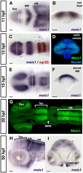
Developmental time course of meis1 mRNA and protein expression. (A, B) mRNA in situ hybridizations (ISHs) for meis1 at 11 hpf showing expression in the eye field, and in the presumptive midbrain (MB) and hindbrain (HB). The transverse section (B) shows meis1 expression in the eye field. (C) mRNA ISH showing meis1 expression at 13 hpf in the optic vesicles, midbrain and hindbrain. egr2b/krox20 expression (red) marks rhombomeres 3 and 5 of the hindbrain. (D) Transverse section of a whole mount immunostain for Meis1 showing protein in the dorsal and ventral leaflets of the 13 hpf optic vesicle. Hoechst 33258 stain marks the nuclei. (E, F) mRNA ISH at 15 hpf showing continued expression of meis1 in the optic vesicles, midbrain and hindbrain. The transverse section (F) shows meis1 expression in the dorsal midbrain. (G) Whole mount immunostain for Meis1 protein at 20 hpf. Meis1 protein is present in the eye, presumptive tectum (Tec), and in the hindbrain (HB) up to the r1-r2 boundary. Meis1 is excluded from the midbrain-hindbrain boundary (MHB). (H, I) mRNA ISH at 50 hpf showing meis1 expression in the hindbrain (HB) and cerebellum (CB) and tectum (Tec). The transverse section (I) shows meis1 expression in the ciliary marginal zone (CMZ) of the retina and in the dorsal midline and a deeper layer of the tectum (white arrow). Embryos in (A, C, E, G) are shown in dorsal view with anterior to the left. Embryo in (H) is shown in lateral view with anterior to the left. Transverse sections in (B, D, F, I) are oriented dorsal up. The dotted lines in (A, C, E, H) indicate the position of the corresponding transverse sections in (B, D, F, I). All scale bars = 50 μm.
|
