
FIGURE
Fig. S3
- ID
- ZDB-FIG-101011-26
- Publication
- Erickson et al., 2010 - Meis1 specifies positional information in the retina and tectum to organize the zebrafish visual system
- Other Figures
- All Figure Page
- Back to All Figure Page
Fig. S3
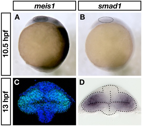
meis1 and smad1 expression in the early optic vesicle. (A, B) mRNA in situ hybridizations for meis1 (A) and smad1 (B) in 10.5-hpf wild-type embryos. The dotted circles indicate the eye fields. Views are lateral with anterior on the top. (C, D) Transverse sections of wild-type 13-hpf optic vesicles stained for Meis1 protein (C) and smad1 mRNA (D). Note that (C) is the same as shown in Figure 1D. The dotted lines outline the optic vesicle and neural tube. Sections are oriented with dorsal at the top. |
Expression Data
Expression Detail
Antibody Labeling
Phenotype Data
Phenotype Detail
Acknowledgments
This image is the copyrighted work of the attributed author or publisher, and
ZFIN has permission only to display this image to its users.
Additional permissions should be obtained from the applicable author or publisher of the image.
Full text @ Neural Dev.