|
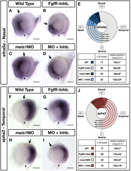
The contribution of Fgf signalling to the NT patterning defects in Meis1-depleted embryos. (A-D, F-I) mRNA in situ hybridizations for the NT markers efna5a (A-D) and epha3 (F-I) in wild type, Meis1-depleted (meis1MO), Fgf receptor-inhibitor treated (FgfR-Inhb.), and FgfR-inhibited/Meis1-depleted retinas (MO + Inhb.). Arrows indicate the extent of the gene expression domain, while the arrowheads indicate the position of the ventral choroid fissure. Representative dissected eyes are shown oriented with dorsal up and nasal to the left. Scale bars = 50 μm. (E, J) Quantification of the changes in efna5a and epha3 expression, as quantified by measuring a 360° profile of in situ staining intensity and graphing the mean radial position at which gene expression intensity falls to the halfway point between its minimum and maximum values. The nmax/2 and tmax/2 values are given as the mean radial position in degrees ± one standard deviation. WT, wild type
|