
Fig. S1
- ID
- ZDB-FIG-100723-21
- Publication
- Iida et al., 2010 - Metalloprotease-dependent onset of blood circulation in zebrafish
- Other Figures
- All Figure Page
- Back to All Figure Page
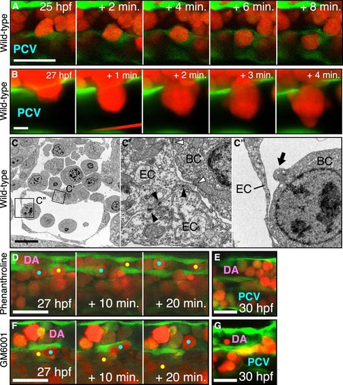
Confocal Images of Erythroid Cells Invading into the Vasculature This figure relates to Figure 1 and 2. (A and B) Time-lapse imagings of their invasion into the PCV in a wild-type embryo. In (A), endothelial cells transiently encapsulate an erythroid cell invading into the PCV. Scale bar represents 25 μm. In (B), an erythroid cell adheres to the PCV with protrusions after invasion. Scale bar represents 5 μm. (C) An electron micrograph of showing adherence between the endothelial cells and between the erythroid and endothelial cells (C′) and a tiny membrane protrusion (black arrow in C″). Scale bar represents 10 μm. (D-G) Confocal images of live embryos injected with ortho-phenanthroline (D and E) or GM6001 (F and G) and showing normal invasion of gata1:mRFP+ cells (color dots) into the vasculature, but a dramatic accumulation inside the blood vessels in the trunk in those embryos. Scale bar represents 25 μm. |