
Fig. 6
- ID
- ZDB-FIG-100616-32
- Publication
- Ito et al., 2010 - Characterization of neural stem cells and their progeny in the adult zebrafish optic tectum
- Other Figures
- All Figure Page
- Back to All Figure Page
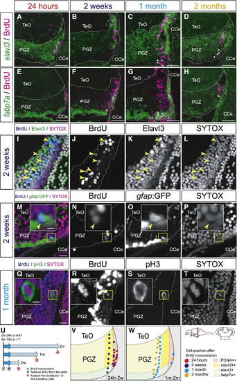
Neural stem/progenitor cells in the mitotic region of the PGZ generate both neuronal and glial cell lineages. (A–H) Distributions of BrdU-positive cells in the elavl3-positive superficial layer (A–D), and the fabp7a-positive deep layer (E–H) of the PGZ in the adult zebrafish optic tectum at 24 h (A, E), 2 weeks (B, F), 1 month (C, G), and 2 months (D, H) post-BrdU administration (14 μm transverse sections, stacked images, dorsal top). After 2 weeks post-BrdU administration, most of the BrdU-positive cells express elavl3 (B–D), however, some BrdU-positive cells express fabp7a but not elavl3 (C, D, G, H, arrowheads). (I–P) Expression of Elavl3 (I–L) and gfap:GFP (M–P) in the BrdU-positive cells at 2 weeks post-BrdU administration (60 μm transverse sections, single planes, dorsal top). Insets in M–P show magnified views of the yellow-boxed areas. The majority of BrdU-positive cells expressed the neuronal marker Elavl3 (I–L), and some BrdU-positive cells expressed the glial maker gfap:GFP (M–P, insets, arrowheads). (Q–T) Distribution of pH3-positive cells in the PGZ at 1 month post-BrdU administration (60 μm transverse sections, single planes, dorsal top). Insets in Q–T show magnified views of the yellow-boxed areas. At 1 month post-BrdU administration, majority of BrdU-positive cells leave the dorsomedial margin (Q, R), while BrdU-positive cells facing the ventricle still undergo cell division and expresses the M-phase marker, pH3 (Q–T, insets). (U) BrdU pulse labeling scheme. (V, W) Summaries of the distribution of BrdU-positive cells at 24 h to 2 weeks (V), and at 1 month to 2 months (W) post-BrdU administration. CCe, corpus cerebelli; PGZ, periventricular gray zone; TeO, tectum opticum. Scale bars: 50 μm in A–H; 10 μm in I, M, Q; 3 μm in inset of M, Q. |
Reprinted from Developmental Biology, 342(1), Ito, Y., Tanaka, H., Okamoto, H., and Ohshima, T., Characterization of neural stem cells and their progeny in the adult zebrafish optic tectum, 26-38, Copyright (2010) with permission from Elsevier. Full text @ Dev. Biol.