
FIGURE
Fig. 4
- ID
- ZDB-FIG-100616-29
- Publication
- Ito et al., 2010 - Characterization of neural stem cells and their progeny in the adult zebrafish optic tectum
- Other Figures
- All Figure Page
- Back to All Figure Page
Fig. 4
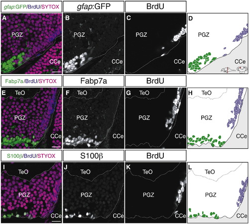
Proliferating cells in the PGZ do not express glial markers. (A–L) Expression of gfap:GFP (A–D), Fabp7a (E–H), and S100β (I–L) in the PGZ of the adult zebrafish optic tectum (60 μm transverse sections, single planes, dorsal top). Proliferating cells are labeled after 72 h of BrdU administration. In the medial region of the PGZ, BrdU-positive proliferating cells (blue) do not express the glial markers, gfap:GFP, Fabp7a and S100β (green); these glial markers are expressed in the deep layer cells. CCe, corpus cerebelli; PGZ, periventricular gray zone; TeO, tectum opticum. Scale bars: 10 μm. |
Expression Data
Expression Detail
Antibody Labeling
Phenotype Data
Phenotype Detail
Acknowledgments
This image is the copyrighted work of the attributed author or publisher, and
ZFIN has permission only to display this image to its users.
Additional permissions should be obtained from the applicable author or publisher of the image.
Reprinted from Developmental Biology, 342(1), Ito, Y., Tanaka, H., Okamoto, H., and Ohshima, T., Characterization of neural stem cells and their progeny in the adult zebrafish optic tectum, 26-38, Copyright (2010) with permission from Elsevier. Full text @ Dev. Biol.