
Fig. 3
- ID
- ZDB-FIG-100616-28
- Publication
- Ito et al., 2010 - Characterization of neural stem cells and their progeny in the adult zebrafish optic tectum
- Other Figures
- All Figure Page
- Back to All Figure Page
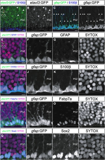
Cells constitute the deep layer of the PGZ are radial glia. (A–D) Distribution of elavl3:GFP-positive cells (A, B) and gfap:GFP-positive cells (C, D) in the PGZ of the adult zebrafish optic tectum (60 μm transverse sections, dorsal top). (A, B) The elavl3:GFP-positive cells are distributed through a broad area of the PGZ, except for the S100β-positive ventral edge. Only a few cells express strong GFP signal and extend dendrite-like processes toward the surface layers of the optic tectum (arrowheads). (C, D) The gfap:GFP-positive cells are distributed along the ventral edge of the PGZ, and extend radial fibers (arrowheads). (E–T) Magnified views of the gfap:GFP-positive cells along the ventral edge of the PGZ (60 μm transverse section, dorsal top). The gfap:GFP-positive cells constitute the deep layer of the PGZ. These cells show immunoreactivities with glial markers, such as GFAP, S100β and Fabp7a (E–P), and the neural stem/progenitor marker Sox2 (Q–T). Scale bars: 10 μm in A, C; 5 μm in E, I, M, Q. |
Reprinted from Developmental Biology, 342(1), Ito, Y., Tanaka, H., Okamoto, H., and Ohshima, T., Characterization of neural stem cells and their progeny in the adult zebrafish optic tectum, 26-38, Copyright (2010) with permission from Elsevier. Full text @ Dev. Biol.