
Fig. S1
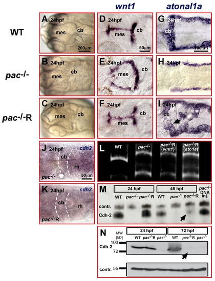
Temporal rescue of pac-/- embryos is restricted to developmental stages prior to cerebellar GC migration. (A–I) Dorsal views of zebrafish embryonic heads at 24 hpf. (A–C) Light microscopy of cerebellar primordium in WT (A) and pac-/- (B) embryos that can be rescued by Cdh2-mRNA injection in pac-/-R-embryos (C). (D–I) This rescue is confirmed by ISH showing the reconstitution of wnt1 dorsal midline expression (D–F) and atonal1a expression throughout the rhombic lip (G–I) in pac-/-R embryos. (L) RT-PCR confirms pac-/- genotype in rescued mutant embryos (tail clip RT-PCR lanes 3+4 was performed on same embryos displayed in F and I, respectively). (J, K) Whole-mount ISH analysis of cadherin-2 expression reveals that cadherin-2 mRNA is hardly detectable in both pac-/- (J) and pac-/-R-embryos (K) at 24 hpf (dorsal views of hindbrain). (M, N) In contrast, Western blot analysis of total embryo extracts including the membrane fractions detects Cadherin-2 protein in pac-/-R-embryos at 24 hpf, with levels comparable to WT embryos (lane 1). By 48 hpf, at the onset of GC migration, Cadherin-2 protein is mostly degraded in pac-/-R-embryos (M, lane 6, black arrow) and completely absent at 72 hpf (N, lane 6). While Cdh2 protein derived from mRNA injections is mostly degraded at 48 hpf, Cdh-2 protein is continuously expressed from plasmid DNA (M, lane 7). Loading controls: TenascinR in (M) and β-Tubulin in (N). cb, cerebellum; mes, mesencephalon; rh, rhombencephalon. |