
FIGURE
Fig. 10
Fig. 10
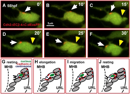
In migrating GCs Cadherin-2 is relocalized along the membrane. (A–F) Time-lapse sequence of Cdh2ΔEC2-4ΔC:dEosFP clusters, UV-converted at the membrane of a migrating GC in a gata1:GFP embryo (white arrowheads). A photo-converted cluster (red) moves successively toward the leading edge during GC elongation and migration (yellow arrowheads mark the trailing edge of the cell, see also Video S15). (G–J) Model of Cdh2-regulated granule cell migration (see text). MHB, midbrain-hindbrain boundary; URL, upper rhombic lip. |
Expression Data
Expression Detail
Antibody Labeling
Phenotype Data
Phenotype Detail
Acknowledgments
This image is the copyrighted work of the attributed author or publisher, and
ZFIN has permission only to display this image to its users.
Additional permissions should be obtained from the applicable author or publisher of the image.
Full text @ PLoS Biol.