
Fig. 9
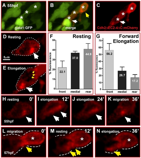
Cadherin-2 relocalization is coordinated with GC migration. (A–E, H–N) Single optical sections of time-lapse recordings using laser-scanning confocal microscopy. (A–C) Heat shock-induced expression of Cadherin-2 reporter protein (Cdh2ΔC2-4ΔC:mCherry) in a cerebellar GC chain (white asterisks). White arrows in (B, C) mark the neuron shown in (H–K), see also Video S14. (D, E) Cdh2ΔC2-4ΔC:mCherry fluorescence is localized in clusters in contact with neighboring GCs (E, yellow arrowheads), but cluster localization changes during migration mostly differing during resting (D) and forward elongation (E) of GCs prior to migration. (F) Quantification of mCherry-fluorescence shows that in resting GCs Cadherin-2 clusters are more evenly distributed, but preferentially localized in the medial and rear compartment of the cell (n = 5, p = 0.056, error bars indicate SEM). (G) Forward migration, in contrast, leads to Cadherin-2 redistribution towards the front compartment of GCs (n = 5, p<0.01). (H–K) Individual images of a time-lapse movie demonstrating the relocation of the Cdh2ΔC2-4ΔC:mCherry reporter (white arrow) after a resting phase from the rear of the cell (H) along the cytoplasmic membrane (I, J) during elongation of the GC to the front (K) to prepare for the next forward movement. (L–N) Localization of Cdh2ΔEC2-4ΔC-mCherry reporter in GCs contacting each other in a migratory GC chain (B, yellow arrowhead). (L) Fluorescence is condensed at contact sites of GCs but partly being redistributed according to migratory movements (M, N). |