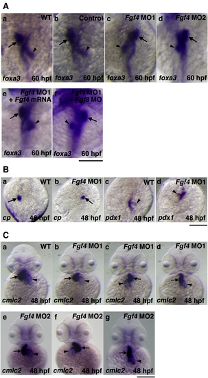
The effect of Fgf4 knockdown on left–right patterning. (A) The expression of foxa3, a marker gene for endoderm, was examined by whole mount in situ hybridization using a digoxigenin-labeled antisense foxa3 cRNA probe. The expression of foxa3 showed that the liver and pancreas were positioned to the left and right of the midline in wild-type embryos, respectively at 60 hpf (94.1%, n = 34) (a). Control MO-injected embryos also showed similar LR patterning of the liver and pancreas (93.8%, n = 32) (b). In contrast, the positions of these organs were randomized in Fgf4 MO1-injected embryos (normal (61.3%, n = 31); reversed (38.7%, n = 31)) (c). The expression of foxa3 showed similar randomized positioning in Fgf4 MO2-injected embryos (normal (56.7%, n = 30); reversed (43.3%, n = 30)) (d). The injection of Fgf4 mRNA significantly rescued the defects in Fgf4 MO1-injected embryos (80.6%, n = 31) (e). The injection of both Fgf4 MO1 and Fgf8 MO also revealed randomized positioning (58.1%, n = 31) (f). Arrows and arrowheads indicate the liver and pancreas, respectively. A scale bar = 200 μm. (B) The expression of ceruloplasmin and pdx1, marker genes for the liver and pancreas, respectively, in embryos at 48 hpf was examined. Their expression confirmed randomized positioning of the liver and pancreas (liver, normal (60%, n = 30); reversed (40%, n = 30): pancreas, normal (63.3%, n = 30); reversed (36.7%, n = 30)) in Fgf4 MO1-injected embryos (a–d). Arrows and arrowheads indicate the liver and pancreas, respectively. A scale bar = 200 μm. (C) The expression of cmlc2, a marker gene for the heart, in embryos at 48 hpf was examined. After the heart tube has formed, leftward movement of the heart (jogging) is followed by rightward bending of the ventricle (D-looping) in wild-type embryos (a). In contrast, Fgf4 MO1-injected embryos exhibited randomized looping (D-looping (38.5%, n = 78); reversed looping (L-looping) (47.4%, n = 78); no looping (14.1%, n = 78)) (b–d). Fgf4 MO2-injected embryos also exhibited randomized looping (D-looping (34.2%, n = 38); reversed looping (L-looping) (50%, n = 38); no looping (15.8%, n = 38)) (e–g). Arrows and arrowheads indicate the ventricle and atrium, respectively. A scale bar = 200 μm.
|
