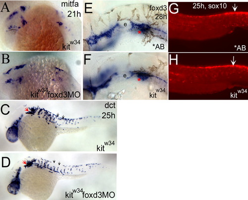
foxd3 loss-of-function does not affect the number of melanoblasts specified from kitw34 neural crest. A,B: mitfa expression is detected in the head and anterior trunk of kitw34 21 hours postfertilization (hpf) embryos. This pattern is not dramatically altered in kitw34 foxd3 morphants. C,D: In 25 hpf kitw34 embryos, dopachrome tautomerase (dct) -positive cells are found caudal to the eye and ear (red arrow), and in streams migrating ventrally between somites from the dorsal trunk (black arrows). The kitw34 foxd3 morphants show some differences in migration (compare C and D, black arrows), but the overall number of cells appears similar. E,F: At 28 hpf, AB and kitw34 embryos, processed by in situ hybridization for foxd3 message. foxd3 expression appears unchanged by kit loss-of-function. E: The developing ear or otic vesicle. G,H: At 25 hpf, AB and kitw34 embryos, processed by fluorescent in situ hybridization for sox10 message. sox10 expression in premigratory neural crest cells is mostly unchanged (white arrows), although there is some reduction in peripheral expression, consistent with the reduction in melanoblast migration observed in kitw34 mutants.
|
