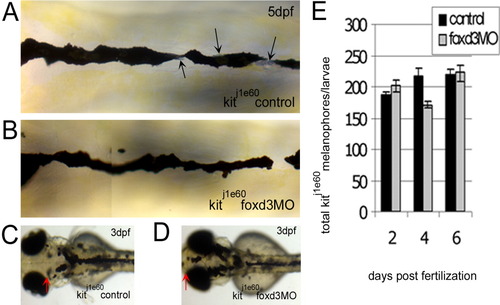
The increase in kit mutant melanophores with foxd3 loss-of-function is not due to transfating of other foxd3-dependent cell types. A-D: The kitj1e60 melanophore migration mutants were injected (or left uninjected; control) with foxd3 morpholino oligonucleotide (MO; foxd3MO), imaged at 3 (C,D) and 5 days postfertilization (dpf; A,B) and fixed at 2, 4, and 6 dpf for melanophore quantification. A: Melanophores localized to the anterior dorsal trunk display relatively normal, nonapoptotic morphology. Black arrows indicate silver pigment cells, iridophores. B: In kitj1e60 foxd3 morphants, foxd3-dependent iridophores are gone indicating MO function (average control and morphant iridophores ± standard deviation at 4 dpf are 52±3 and 18±14, respectively), yet melanophore morphology and number remain similar to uninjected controls. C: The kitj1e60 control melanophores do not localize to the anterior most portion of the head, indicating a migration defect. D: The presence of melanophores over the head is partially rescued with foxd3 loss-of-function (red arrows indicate anterior extent of melanophores). E: The kitj1e60 melanophores are not significantly increased with foxd3 loss-of-function at 2, 4, and 6 dpf (P = 0.31 by 2-way analysis of variance; 8-15 fish per time point).
|
