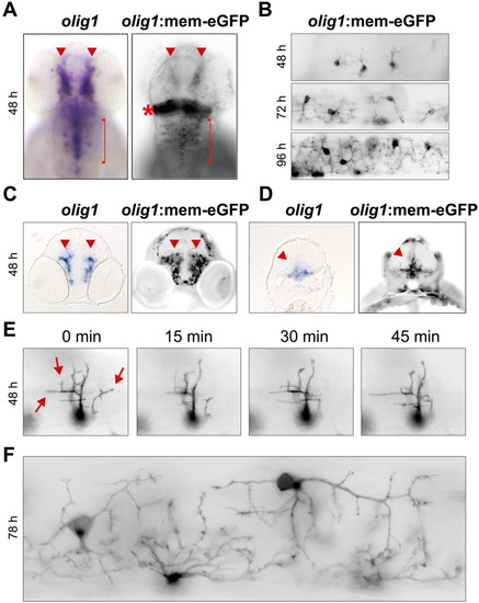
The olig1 promoter is sufficient to drive membrane-targeted eGFP expression in developing oligodendrocytes. A: eGFP expression from the 5.4-kb zebrafish olig1 promoter recapitulates endogenous olig1 expression determined by in situ hybridization. Red arrowheads indicate midbrain oligodendrocytes and red bracket demarcates hindbrain oligodendrocytes. Red asterisk marks ectopic expression of eGFP in the region of the cerebellum. B: Developmental time course demonstrates increasingly complex network of oligodendrocyte processes in the spinal cord of olig1:mem-eGFP transgenic animals at 48, 72, and 96 hpf. C,D: Transverse sections of olig1 in situ hybridizations and eGFP fluorescence at the level of the fore-midbrain (C) and hindbrain (D). Red arrowheads indicate olig1 expression domains. E: Time-lapse microscopy images of an individual oligodendrocyte in the 48-hpf spinal cord demonstrating its highly motile, extending, and retracting, membrane processes (red arrows). F: Close-up of eGFP+ oligodendrocytes in the spinal cord of a 78-hpf transgenic animal.
|
