
Fig. 6
- ID
- ZDB-FIG-090224-51
- Publication
- Dowling et al., 2009 - Loss of myotubularin function results in T-tubule disorganization in zebrafish and human myotubular myopathy
- Other Figures
- All Figure Page
- Back to All Figure Page
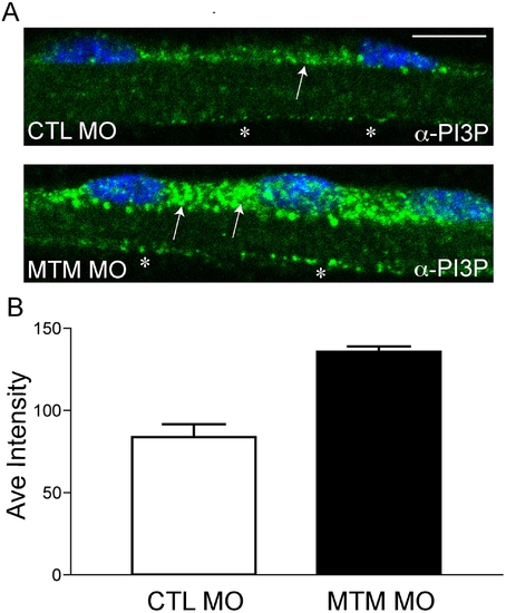
Increased PI3P levels in myotubularin morphant myofibers. (A) Representative myofibers immunostained with anti-PI3P. Perinuclear staining of PI3P in myotubularin morphant myofibers is much more abundant than in control myofibers (arrows). There is also a modest increase in membrane localized PI3P (*). Scale bar = 10 mm. (B) Quantitation of PI3P immunofluorescence. PI3P intensity measured over a uniform perinuclear area (see methods for details) and was 83.7+/-7.8 pixels for control morphants and 135.5+/-3.3 for myotubularin morphants (3 trials; p = 0.0027). This represented a 1.6x increase in PI3P staining intensity. |
| Fish: | |
|---|---|
| Knockdown Reagent: | |
| Observed In: | |
| Stage: | Protruding-mouth |