
Fig. 4
- ID
- ZDB-FIG-090224-49
- Publication
- Dowling et al., 2009 - Loss of myotubularin function results in T-tubule disorganization in zebrafish and human myotubular myopathy
- Other Figures
- All Figure Page
- Back to All Figure Page
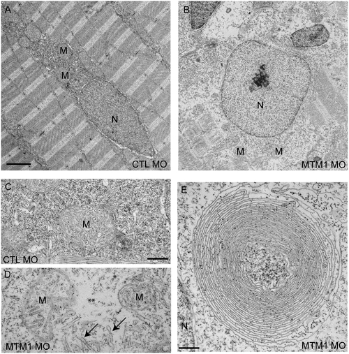
Abnormal perinuclear ultrastructure in 72 hpf myotubularin morphants. Comparison of the perinuclear area from control (CTL MO) and myotubularin (MTM MO) morphants. (A, B) Control injected embryos had thin myonuclei (N) with well organized perinuclear organelles (M = mitochondria). Myotubularin injected embryos had large, rounded nuclei (N) and disorganized perinuclear compartments. Three embryos from three independent injections were examined. Higher magnification (C, D) of the perinuclear compartment revealed abnormal mitochondria (M), areas nearly devoid of organelles (**), and several tubule-like structures (arrows). (E) Example of an unusual membranous perinuclear structure. Such structures were observed in multiple myofibers. Scale bars: A, B (2 mm), C–E (500 nm). |
| Fish: | |
|---|---|
| Knockdown Reagent: | |
| Observed In: | |
| Stage: | Protruding-mouth |