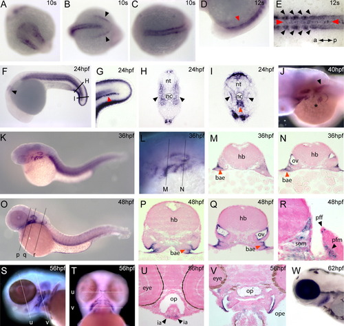
Expression of frem3 during development. frem3 is broadly expressed during embryonic development in zebrafish. A-C: Expression is first detected during early somitogenesis and, at the bud stage is detected in the lateral head mesoderm (A, anterior), somites (B, posterior; C, dorsal), and differentiating tail ectoderm (B, arrowhead). E,I: In newly differentiating caudal somites, expression is highest in the adaxial cells adjacent to the neural tube (black arrowheads). F,H: By 24 hpf expression is detected throughout the somitic musculature (F, lateral; H, in section, arrowhead). J: Expression is noted in the developing pectoral fin musculature (asterisk) which persists in the adjacent somites 1 and 2 (arrowhead). F-I: As with the other frem-related genes, expression is detected in the caudal fin folds. D,E,G,I: In addition to somitic expression, transcripts are observed in the hypochord (red arrowheads) as a single medial line of cells. K-R: During branchial arch development frem3 is expressed in the endodermal pouches (K-O; in section P,Q,R, red arrowheads). Expression is also noted in the otic placode (F, arrow) and by later developmental stages this expression is highest in the dorsolateral aspect of the otic vesicle (N,Q). S-V: Expression is initiated in the musculature of the developing head and in the hyoid posterior ectodermal margin and extending into the medial ectoderm (S-U, and in section, V). W: Expression in the operculum persists during later embryogenesis. nt, neural tube; nc, notochord; hb, hindbrain; bae, branchial arch endoderm; ov, otic vesicle; pff, pectoral fin fold; som, somite; pfm, pectoral fin muscle; op, oropharynx; ia, intermandibularis posterior muscle; ope, operculum.
|
