
Fig. 4
- ID
- ZDB-FIG-080925-27
- Publication
- Lee et al., 1999 - A wave of free cytosolic calcium traverses zebrafish eggs on activation
- Other Figures
- All Figure Page
- Back to All Figure Page
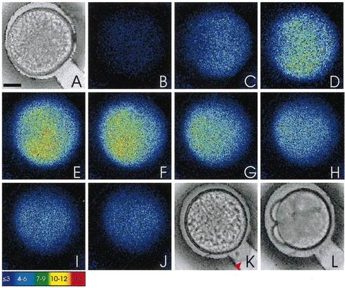
Representative sequence of images from an aequorin-loaded egg illustrating changes in intracellular free calcium during activation in the presence of sperm. A is a bright-field image of the unactivated egg just prior to the initiation of the signal and once again shows no separation between the chorion and the egg plasma membrane, whereas K shows the egg after the passage of the calcium wave, clearly indicating (see arrowhead) a raised chorion. L shows the egg at the 4-cell stage, indicating that it has been fertilized and is developing normally. The photon images (B to J) represent 60 s of accumulated light, with a 20-s step separating each successive image. The dividing blastodisc in L indicates the location of the AP. Color scale indicates luminescent flux in photons per pixel. Scale bar is 200 μm. |
Reprinted from Developmental Biology, 214(1), Lee, K.W., Webb, S.E., and Miller, A.L., A wave of free cytosolic calcium traverses zebrafish eggs on activation, 168-180, Copyright (1999) with permission from Elsevier. Full text @ Dev. Biol.