
|
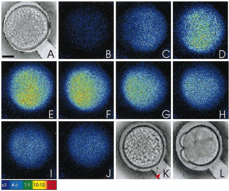
Fig. 4 Representative sequence of images from an aequorin-loaded egg illustrating changes in intracellular free calcium during activation in the presence of sperm. A is a bright-field image of the unactivated egg just prior to the initiation of the signal and once again shows no separation between the chorion and the egg plasma membrane, whereas K shows the egg after the passage of the calcium wave, clearly indicating (see arrowhead) a raised chorion. L shows the egg at the 4-cell stage, indicating that it has been fertilized and is developing normally. The photon images (B to J) represent 60 s of accumulated light, with a 20-s step separating each successive image. The dividing blastodisc in L indicates the location of the AP. Color scale indicates luminescent flux in photons per pixel. Scale bar is 200 μm.
Reprinted from Developmental Biology, 214(1), Lee, K.W., Webb, S.E., and Miller, A.L., A wave of free cytosolic calcium traverses zebrafish eggs on activation, 168-180, Copyright (1999) with permission from Elsevier. Full text @ Dev. Biol.