
Fig. 7
- ID
- ZDB-FIG-080925-29
- Publication
- Lee et al., 1999 - A wave of free cytosolic calcium traverses zebrafish eggs on activation
- Other Figures
- All Figure Page
- Back to All Figure Page
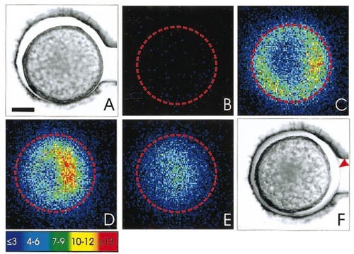
Representative series of images of a vegetal pole (VP) view of an activation wave indicating that the wave propagates mainly in the periphery of the egg. A and F are bright-field images of the egg before and after activation, respectively. The arrowhead in F indicates the raised chorion. B illustrates the resting level of luminescence prior to activation. C indicates the activation wave at the equator of the egg, and D and E demonstrate the subsequent progression of the wave to the VP. The location of the egg equator is outlined. B to E show 60 s of accumulated light. Color scale indicates luminescent flux in photons per pixel. Scale bar is 200 μm. |
Reprinted from Developmental Biology, 214(1), Lee, K.W., Webb, S.E., and Miller, A.L., A wave of free cytosolic calcium traverses zebrafish eggs on activation, 168-180, Copyright (1999) with permission from Elsevier. Full text @ Dev. Biol.