
FIGURE
Fig. S5
- ID
- ZDB-FIG-080829-57
- Publication
- Kishi et al., 2008 - The identification of zebrafish mutants showing alterations in senescence-associated biomarkers
- Other Figures
- All Figure Page
- Back to All Figure Page
Fig. S5
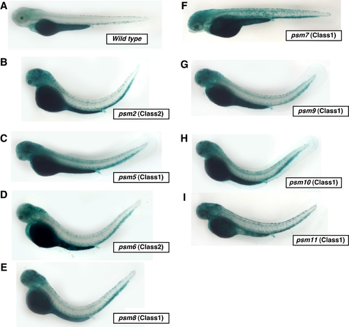
Homozygous psm mutant embryos showing high levels of tissue-specific SA-β-gal activity without oxidative stress. Homozygous 3.5 dpf neurodegenerative zebrafish psm mutants (psm2, 5, 6, 8, 9, 10, 11) show much higher levels of punctate SA-β-gal staining throughout their central nervous system compared with wild-type embryos (with PTU). Of note, the muscle atrophy mutant psm7 shows particularly high levels of punctate staining in the trunk region. |
Expression Data
Expression Detail
Antibody Labeling
Phenotype Data
Phenotype Detail
Acknowledgments
This image is the copyrighted work of the attributed author or publisher, and
ZFIN has permission only to display this image to its users.
Additional permissions should be obtained from the applicable author or publisher of the image.
Full text @ PLoS Genet.