Senescence, cell death and ROS generation in homozygous psm zebrafish mutants in the absence of oxidative stress.
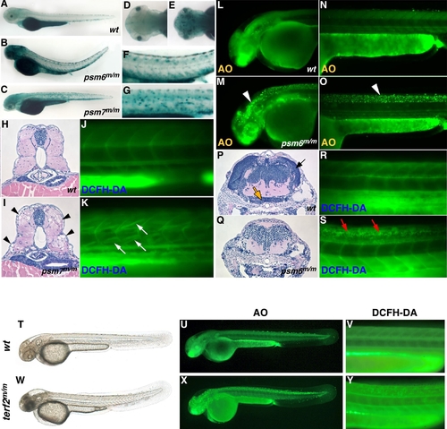
SA-β-gal activity was found to be high throughout the brain and neural tube in neurodegenerative zebrafish mutants (B and the magnified trunk region in F) [psm6m/m is shown] compared with wild-type embryos (A) at 3.5 dpf (with PTU). (D, E) Dorsal views of the head of wild-type and psm6m/m embryos respectively. (G) Neurodegenerative mutant embryos have high levels of acridine orange (AO) staining (white arrowheads) in the brain (M) and neural tube (O), compared with wild-type embryos (L, N) at 2 dpf. H&E staining of transverse sections of the head of 5-day old (5 dpf) larvae reveals a reduction in the number of neuronal nuclei and absence of brain structures in neurodegenerative mutants (Q), compared with wild-type embryos (arrows in P which indicate the tectum opticum [black arrow] and caudal hypothalamus [orange arrow]). DCFH-DA staining indicates high ROS generation in the neural tube of neurodegenerative mutants (red arrows in S) compared with wild-type embryos (R) at 3.5 dpf. The muscle atrophy mutant [psm7m/m is shown] is characterized by punctate SA-β-gal activity in the trunk (C and magnified trunk region in G). H&E staining of transverse sections of the trunks of 4-day old (4 dpf) psm7m/m larvae reveals the loss of muscle fibers (black arrowheads in I), compared with wild-type embryos (H). DCFH-DA staining reveals the generation of ROS in skeletal muscle in the absence of exogenous oxidative stress in psm7m/m embryos (white arrows in K) compared with wild-type embryos (J) at 3.5 dpf. Morphology of terf2m/m mutant embryo was compared with that of wild-type sibling (T, W) at 2 dpf (with PTU). terf2m/m mutant embryos have high levels of acridine orange (AO) staining (X) in the brain and neural tube, compared with wild-type embryos (U) at 2 dpf. DCFH-DA staining indicates high ROS generation in the neural tube of terf2m/m mutant (Y) compared with wild-type embryos (V) at 3.5 dpf.
|
