
Fig. 3
- ID
- ZDB-FIG-080514-9
- Publication
- Köster et al., 2001 - Tracing transgene expression in living zebrafish embryos
- Other Figures
- All Figure Page
- Back to All Figure Page
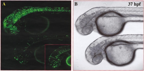
Combined Gal4-VP16-activator/effector constructs result in strong EGFP expression in numerous cells of DNA-injected embryos. Single optical confocal microscopy section (lateral view) focusing on the nervous system of injected embryos at 37 hpf. Two embryos were embedded together in low-melting agarose to allow direct comparison of EGFP-expression levels and number of EGFP-expressing cells in a single optical section. Fluorescent (A pseudocolored in green) and transmitted light (B) images were taken at the same focal plane without moving the embryos. (A, B) EF-GVP-UG-injected embryos of the same batch. EGFP expression in EF-GVP-UG-injected embryos can vary strongly in intensity and frequency from a high number of embryonic GFP-expressing cells (A, upper embryo) to very few EGFP-expressing cells (A, lower embryo). Higher laser power and increased contrast, however, revealed a higher frequency of EGFP-expressing cells in the lower embryo than judged by the initial conditions (see A, inset). |
Reprinted from Developmental Biology, 233(2), Köster, R.W. and Fraser, S.E., Tracing transgene expression in living zebrafish embryos, 329-346, Copyright (2001) with permission from Elsevier. Full text @ Dev. Biol.