
Fig. 9
- ID
- ZDB-FIG-080514-14
- Publication
- Köster et al., 2001 - Tracing transgene expression in living zebrafish embryos
- Other Figures
- All Figure Page
- Back to All Figure Page
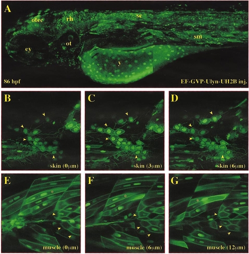
Gal4-VP16 is capable of mediating coexpression of two UAS-dependent transgenes in individual cells of living zebrafish embryos. (A–G) Single optical sections through EF-GVP-Ulyn-UH2B-injected embryo at 86 hpf taken by confocal microscopy (EGFP fluorescence pseudocolored in green). (A) Lateral view of EF-GVP-Ulyn-UH2B-injected embryo, the high frequency of EGPF-expressing cells allows one to observe the overall morphology and to distinguish different tissues like nervous system, body muscles, and yolk syncytial layer. (B–G) Individual optical sections of stacks of pictures taken from the skin (B–D) or somitic muscles (E–G) of the injected embryo. Arrowheads mark same individual cells through these stacks to demonstrate that cells indeed coexpress both EGFP variants while scoring single individual optical sections might often underestimate the degree of coexpression. Note syncytial nature of muscle cells containing several nuclei (E*). Abbreviations: ey, eye; ot, otic vesicle; otec, optic tectum; rh, rhombencephalon; sc, spinal cord; sm, somitic muscles; y, yolk. |
Reprinted from Developmental Biology, 233(2), Köster, R.W. and Fraser, S.E., Tracing transgene expression in living zebrafish embryos, 329-346, Copyright (2001) with permission from Elsevier. Full text @ Dev. Biol.