
Fig. 8
- ID
- ZDB-FIG-080514-13
- Publication
- Köster et al., 2001 - Tracing transgene expression in living zebrafish embryos
- Other Figures
- All Figure Page
- Back to All Figure Page
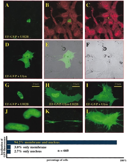
Gal4-VP16-mediates coexpression of two UAS effectors in a very high frequency. (A–L) Confocal microscopy pictures of cultured primary zebrafish cells transfected with different Gal4-VP16 activator/effector constructs. (A–C) EF-GVP/UH2B cotransfected cells, images of H2BEYFP fluorescence (A, pseudocolored in green) and Bodipyceramide-TexasRed counterstained cells (C, pseudocolored in red) were overlaid (B) to confirm nuclear localization of the histone2B-EYFP-fusion protein. (D–F) EF-GVP/Ulyn cotransfected cells, fluorescent (D), and transmitted (F) light images were overlaid (E) to confirm the membrane localization and nuclear exclusion of the lynEGFP-fusion protein. Comparison of EF-GVP/UH2B cotransfected (G, J), EF-GVP/Ulyn cotransfected (I, L), and EF-GVP-Ulyn-UH2B-transfected cells (H, K) shows characteristic localization, size, and shape of the different EGFP-targeted organelles (pseudocolored in green) to allow scoring of simultaneous coexpression of both EGFP variants in individual and EF-GVP-Ulyn-UH2B-transfected cells. Scoring results in percentages are shown in the diagram below. |
Reprinted from Developmental Biology, 233(2), Köster, R.W. and Fraser, S.E., Tracing transgene expression in living zebrafish embryos, 329-346, Copyright (2001) with permission from Elsevier. Full text @ Dev. Biol.