
Fig. 2
- ID
- ZDB-FIG-080514-8
- Publication
- Köster et al., 2001 - Tracing transgene expression in living zebrafish embryos
- Other Figures
- All Figure Page
- Back to All Figure Page
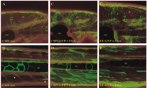
Gal4-VP16-mediated expression in comparison to conventional DNA-construct expression. Lateral views of the hindbrain (A, C, E) and trunk (B, D, F) of CMV-lyn-injected (A, B), CMV-GVP/Ulyn-coinjected, and EF-GVP/Ulyn-coinjected embryos at 3 dpf. Pictures represent pseudocolored composites of single optical sections using confocal microscopy. EGFP-expressing cells are displayed in green. Embryos where counterstained with Bodipyceramide TexasRed (Molecular Probes) displayed in red to visualize overall morphology. Note the precise membranelocalization of the lynEGFP-fluorescence in expressing cells (e.g., neuron in A or muscle cells in D). Abbreviations: nc, notochord; nt, neural tube; otv, otic vesicle; rh, rhombencephalon. |
Reprinted from Developmental Biology, 233(2), Köster, R.W. and Fraser, S.E., Tracing transgene expression in living zebrafish embryos, 329-346, Copyright (2001) with permission from Elsevier. Full text @ Dev. Biol.